In the field of medical testing, the clarity and accuracy of images are directly related to the accuracy of diagnosis and the therapeutic effect. When SONY ILX-LR1, a high-resolution camera specifically designed for drone inspection, enters the medical field, it is quietly triggering a revolution in medical testing.

I. From the Sky to the Operating Table: The Cross-border Journey of ILX-LR1
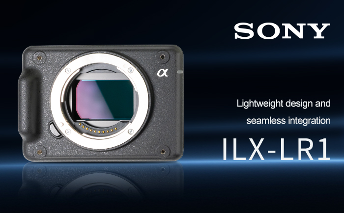
The SONY ILX-LR1 was originally designed as a professional camera for industrial applications such as drone inspection and mapping measurement. It is equipped with a 35mm full-frame back-illuminated Exmor R™ CMOS image sensor with approximately 61 million effective pixels, weighs only about 243 grams, and has a compact body size of 100.0×74.0x42.5mm, which can be easily integrated into various devices.
It is precisely these characteristics that enable it to demonstrate great potential in the field of medical testing. Medical imaging requires capturing subtle lesions and tissue structures, and the high-resolution feature of ILX-LR1 precisely meets this demand.
Ii. The New “Eyes” for Medical Testing: The Breakthrough Advantage of 61 Million Pixels
More detail capture: The 61-megapixel ILX-LR1 offers unprecedented image clarity for medical testing. Even the tiniest lesions or structural changes can be clearly captured, which is of great significance for improving the accuracy of diagnosis and the success rate of surgery. High-resolution images provide doctors with more detailed visual information, enabling every tiny problem and potential hazard to be clearly presented.
Flexible zooming and cropping: Due to the sufficient number of pixels, the images captured by ILX-LR1 can be zoomed in and cropped without losing clarity. This is very helpful for medical tests that require detailed observation of specific areas or structures. For instance, during an endoscopy, doctors can magnify images to examine more closely the minute changes in the intestines or respiratory tract.
Enhanced image analysis capabilities: The 61-megapixel images of ILX-LR1 provide more detailed information, which makes image-based algorithms and artificial intelligence (AI) analysis more effective. In pathology, high-definition images can help AI algorithms identify cancer cells or other abnormal cells more accurately. In radiology, high-pixel images can support more precise three-dimensional reconstruction and more accurate lesion localization.

Iii. New Possibilities of Telemedicine: Collaboration Beyond Geographical Limitations
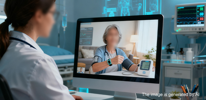
The ILX-LR1 supports remote control and real-time viewfinder monitoring, which enables doctors to conduct efficient medical collaboration at different locations and times. This ability is particularly important in medical diagnosis in remote areas, enabling expert resources to break through geographical limitations and provide precise medical services to more patients.
IV. Lightweight Design and seamless Integration: Suitable for more medical scenarios
The lightweight design (only 243 grams) and compact size of ILX-LR1 make it easy to integrate into various medical devices without increasing the burden on the equipment. All six sides of the camera are equipped with M3 screw holes, allowing users to install the camera on medical testing equipment at different positions, providing diverse installation and usage methods.
The USB Type-C interface and Micro HDMI interface on the back of the camera provide more possibilities for expansion and design in the field of medical testing, which can meet the diverse needs of users. This compatibility enables the ILX-LR1 to be flexibly integrated into various medical devices and systems.

V. Practical Application Scenarios: The Multiple Roles of ILX-LR1 in Medical Testing
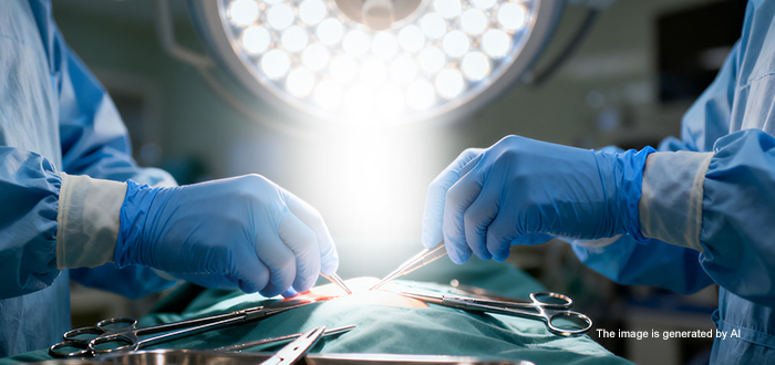
1. Surgical assistance and medical imaging: During the surgical process, doctors can clearly see the details of the surgical site through the high-definition images captured by ILX-LR1, thereby making more accurate judgments and operations. This not only increases the success rate of the surgery but also significantly reduces the surgical risks.
2. Pathological analysis: High-definition images can help AI algorithms more accurately identify cancer cells or other abnormal cells, improving the accuracy and efficiency of pathological diagnosis.
3. Endoscopy: Doctors can magnify the images captured by ILX-LR1 to more closely examine the minute changes in the intestines or respiratory tract, achieving a more accurate diagnosis.
4. Medical Education and Training: High-resolution images and video materials can be used for medical education and training, enabling students and medical staff to have a clearer understanding of human body structure and lesion characteristics.
Conclusion
The SONY ILX-LR1 camera, with its 61-megapixel high pixel, lightweight design and remote control capabilities, is demonstrating outstanding application potential in the medical testing field. It breaks through the limitations of traditional medical imaging, providing doctors with clearer and more accurate visual information and helping medical testing reach new heights.
 Sony FCB camera block